Friday, July 11, 2008

The Structure of the Skin

Skin layers:
1. epidermis
2. dermis
3. subcutis
showing a
a. hair follicle
b. sweat gland
c. sebaceous gland

The skin consists of an outer, protective layer (epidermis) and an inner, living layer (dermis). The top layer of the epidermis is composed of dead cells containing keratin, the horny scleroprotein that also makes up hair and nails.
source: wiki/encarta encyclopedia

The SKIN

In zootomy and dermatology, skin is the largest organ of the integumentary system made up of multiple layers of epithelial tissues that guard underlying muscles and organs.Skin pigmentation varies among populations, and skin type can range from dry skin to oily skin.

The adjective cutaneous literally means "of the skin" (from Latin cutis, skin).
Because it interfaces with the environment, skin plays a very important role in protecting (the body) against pathogens. Its other functions are insulation, temperature regulation, sensation, synthesis of vitamin D, and the protection of vitamin B folates.
Severely damaged skin will try to heal by forming scar tissue. This is often discolored and depigmented


The skin is an organ of double-layered tissue stretched over the surface of the body and protecting it from drying or losing fluid, from harmful external substances, and from extremes of temperature. The inner layer, called the dermis, contains sweat glands, blood vessels, nerve endings (sense receptors), and the bases of hair and nails. The outer layer, the epidermis, is only a few cells thick; it contains pigments, pores, and ducts, and its surface is made of dead cells that it sheds from the body. (Hair and nails are adaptations arising from the dead cells.) The sweat glands excrete waste and cool the body through evaporation of fluid droplets; the blood vessels of the dermis supplement temperature regulation by contracting to preserve body heat and expanding to dissipate it. Separate kinds of receptors convey pressure, temperature, and pain. Fat cells in the dermis insulate the body, and oil glands lubricate the epidermis.
source: wiki/encarta encyclopedia
.

The Male Organ

The organs of the male reproductive system enable a man to have sexual intercourse and to fertilize female sex cells (eggs) with sperm. The gonads, called testicles, produce sperm. Sperm pass through a long duct called the vas deferens to the seminal vesicles, a pair of sacs that lies behind the bladder. These sacs produce seminal fluid, which mixes with sperm to produce semen. Semen leaves the seminal vesicles and travels through the prostate gland, which produces additional secretions that are added to semen. During male orgasm the penis ejaculates semen.

The Reproductive System

The human male reproductive system is a series of organs located outside of the body and around the pelvic region of a male that contribute towards the reproductive process.
The male contributes to reproduction by producing spermatozoa. The spermatozoa then fertilize the egg in the female body and the fertilized egg (zygote) gradually develops into a fetus, which is later born as a child

Reproduction is accomplished by the union of male sperm and the female ovum. In coitus, the male organ ejaculates more than 250 million sperm into the vagina, from which some make their way to the uterus. Ovulation, the release of an egg into the uterus, occurs approximately every 28 days; during the same period the uterus is prepared for the implantation of a fertilized ovum by the action of estrogens. If a male cell fails to unite with a female cell, other hormones cause the uterine wall to slough off during menstruation. From puberty to menopause, the process of ovulation, and preparation, and menstruation is repeated monthly except for periods of pregnancy. The duration of pregnancy is about 280 days. After childbirth, prolactin, a hormone secreted by the pituitary, activates the production of milk.
source: wiki/encarta encyclopedia

The Pituitary Gland

The pituitary gland, or hypophysis, (from Greek hupophuein, to grow up beneath) is an endocrine gland about the size of a pea. It is a protrusion off the bottom of the hypothalamus at the base of the brain, and rests in a small, bony cavity (sella turcica) covered by a dural fold (diaphragma sellae).

The pituitary fossa, in which the pituitary gland sits, is situated in the sphenoid bone in the middle cranial fossa at the base of the brain.

The pituitary gland secretes hormones regulating homeostasis, including trophic hormones that stimulate other endocrine glands. It is functionally connected to the hypothalamus by the median eminence.

The hypophysis is also the top cell of the suspensor in a dicot embryo, which will differentiate to form part of the root cap.

Called the master gland, the pituitary secretes hormones that control the activity of other endocrine glands and regulate various biological processes. Its secretions include growth hormone.
a. (which stimulates cellular activity in bone, cartilage, and other structural tissue); thyroid
stimulating hormone
b. (which causes the thyroid to release metabolism-regulating hormones); antidiuretic hormone
c. (which causes the kidney to excrete less water in the urine); and prolactin
d. (which stimulates milk production and breast development in females).
The pituitary gland is influenced both neurally and hormonally by the hypothalamus.
source:wiki/encarta encyclopedia

The Endocrine System

The endocrine system is an integrated system of small organs that involve the release of extracellular signaling molecules known as hormones. The endocrine system is instrumental in regulating metabolism, growth, development and puberty, tissue function, and also plays a part in determining mood. The field of medicine that deals with disorders of endocrine glands is endocrinology, a branch of the wider field of internal medicine.
In addition to the integrative action of the nervous system, control of various body functions is exerted by the endocrine glands. An important part of this system, the pituitary, lies at the base of the brain.


This master gland secretes a variety of hormones, including the following:
(1)
a hormone that stimulates the thyroid gland and controls its secretion of thyroxine, which dictates the rate at which all cells utilize oxygen;
(2) a hormone that controls the secretion in the adrenal gland of hormones that influence
the metabolism of carbohydrates, sodium, and potassium and control the rate at which substances are exchanged between blood and tissue fluid;
(3) substances that control the secretion in the ovaries of estrogen and progesterone
and the creation in the testicles of testosterone;
(4) the somatotropic, or growth, hormone, which controls the rate of development of
the skeleton and large interior organs through its effect on the metabolism of
proteins and carbohydrates;
(5) an insulin inhibitor—a lack of insulin causes diabetes mellitus.

The posterior lobe of the pituitary secretes vasopressin, which acts on the kidney to control the volume of urine; a lack of vasopressin causes diabetes insipidus, which results in the passing of large volumes of urine.
The posterior lobe also elaborates oxytocin, which causes contraction of smooth muscle in the intestines and small arteries and is used to bring about contractions of the uterus in childbirth.

Other glands in the endocrine system are the pancreas, which secretes insulin, and the parathyroid, which secretes a hormone that regulates the quantity of calcium and phosphorus in the blood.
source: wiki/encarta encyclopedia

The Stomach

Located on the left side of the body, under the diaphragm, the stomach is a muscular, saclike organ that connects the esophagus and small intestine. Its main function is to break down food. Cells in the stomach lining secrete enzymes, hydrochloric acid, and other chemicals to continue the digestive process begun in the mouth and produce mucus to keep these substances from digesting the lining itself.

The Digestive and Excretory System

Digestion is the breaking down of food in the body, into a form that can be absorbed and used or excreted. It is also the process by which the body breaks down food into smaller components that can be absorbed by the blood stream. In mammals, preparation for digestion begins with the cephalic phase in which saliva is produced in the mouth and digestive enzymes are produced in the stomach. Mechanical and chemical digestion begin in the mouth where food is chewed, and mixed with saliva to break down starches. The stomach continues to break food down mechanically and chemically through the churning of the stomach and mixing with enzymes. Absorption occurs in the stomach and gastrointestinal tract, and the process finishes with excretion

The excretory system is an organ system that performs the function of excretion, the bodily process of discharging nitrogeneous wastes. It is responsible for the elimination of the nitrogeneous waste products of metabolism as well as other non-useful nitrogeneous materials. The main components of the excretory system are your two kidneys, two tubes that carry urine called ureters, the bladder, and the urethra.

The energy required for maintenance and proper functioning of the human body is supplied by food. After it is broken into fragments by chewing (see Teeth) and mixed with saliva, digestion begins. The food passes down the gullet into the stomach, where the process is continued by the gastric and intestinal juices. Thereafter, the mixture of food and secretions, called chyme, is pushed down the alimentary canal by peristalsis, rhythmic contractions of the smooth muscle of the gastrointestinal system.

The contractions are initiated by the parasympathetic nervous system; such muscular activity can be inhibited by the sympathetic nervous system. Absorption of nutrients from chyme occurs mainly in the small intestine; unabsorbed food and secretions and waste substances from the liver pass to the large intestines and are expelled as feces. Water and water-soluble substances travel via the bloodstream from the intestines to the kidneys, which absorb all the constituents of the blood plasma except its proteins. The kidneys return most of the water and salts to the body, while excreting other salts and waste products, along with excess water, as urine.
source: wiki/encarta encyclopedia

The Human Lungs

Human Lungs Air travels to the lungs though a series of tubes and airways. The two branches of the trachea, called bronchi, subdivide within the lobes into smaller and smaller air vessels. They terminate in alveoli, tiny air sacs surrounded by capillaries. When the alveoli inflate with inhaled air, oxygen diffuses into the blood in the capillaries to be pumped by the heart to the tissues of the body, and carbon dioxide diffuses out of the blood into the lungs, where it is exhaled.

The Respiratory System

In humans and other mammals, the respiratory system consists of the airways, the lungs, and the respiratory muscles that mediate the movement of air into and out of the body. Within the alveolar system of the lungs, molecules of oxygen and carbon dioxide are passively exchanged, by diffusion, between the gaseous environment and the blood. Thus, the respiratory system facilitates oxygenation of the blood with a concomitant removal of carbon dioxide and other gaseous metabolic wastes from the circulation. The system also helps to maintain the acid-base balance of the body through the efficient removal of carbon dioxide from the blood.
Respiration is carried on by the expansion and contraction of the lungs; the process and the rate at which it proceeds are controlled by a nervous center in the brain.
In the lungs, oxygen enters tiny capillaries, where it combines with hemoglobin in the red blood cells and is carried to the tissues. Simultaneously, carbon dioxide, which entered the blood in its passages through the tissues, passes through capillaries into the air contained within the lungs. Inhaling draws into the lungs air that is higher in oxygen and lower in carbon dioxide; exhaling forces from the lungs air that is high in carbon dioxide and low in oxygen. Changes in the size and gross capacity of the chest are controlled by contractions of the diaphragm and of the muscles between the ribs.
source: wiki/encarta encyclopedia

The Immune System


A scanning electron microscope image (photo)
single neutrophil (yellow)
engulfing anthrax bacteria (orange).

An immune system is a collection of mechanisms within an organism that protects against disease by identifying and killing pathogens and tumor cells. It detects a wide variety of agents, from viruses to parasitic worms, and needs to distinguish them from the organism's own healthy cells and tissues in order to function properly. Detection is complicated as pathogens adapt and evolve new ways to successfully infect the host organism.

To survive this challenge, multiple mechanisms evolved that recognize and neutralize pathogens. Even simple unicellular organisms such as bacteria possess enzyme systems that protect against viral infections. Other basic immune mechanisms evolved in ancient eukaryotes and remain in their modern descendants, such as plants, fish, reptiles, and insects. These mechanisms include antimicrobial peptides called defensins, phagocytosis, and the complement system. More sophisticated mechanisms, however, developed relatively recently, with the evolution of vertebrates.

The immune systems of vertebrates such as humans consist of many types of proteins, cells, organs, and tissues, which interact in an elaborate and dynamic network. As part of this more complex immune response, the vertebrate system adapts over time to recognize particular pathogens more efficiently. The adaptation process creates immunological memories and allows even more effective protection during future encounters with these pathogens. This process of acquired immunity is the basis of vaccination.
Disorders in the immune system can result in disease. Immunodeficiency diseases occur when the immune system is less active than normal, resulting in recurring and life-threatening infections. Immunodeficiency can either be the result of a genetic disease, such as severe combined immunodeficiency, or be produced by pharmaceuticals or an infection, such as the acquired immune deficiency syndrome (AIDS) that is caused by the retrovirus HIV. In contrast, autoimmune diseases result from a hyperactive immune system attacking normal tissues as if they were foreign organisms. Common autoimmune diseases include rheumatoid arthritis, diabetes mellitus type 1 and lupus erythematosus. These critical roles of immunology in health and disease are areas of intense scientific study.
source: wikipedia encyclopedia

The NERVOUS System Organization

The Peripheral Nervous System
1.) Sensory Neurons-(Afferent)
2.) Motor Neurons-(efferent)
2a. Autonomic nervous system
2b. Somatic nervous system
b1.) Sympathetic nervous system
b2.) Parasympathetic Nervous system

The Central Nervous System
a.) Brain
b.) Spinal Cord

Thursday, July 10, 2008

The Immune System

Immune System
The body defends itself against foreign proteins and infectious microorganisms by means of a complex dual system that depends on recognizing a portion of the surface pattern of the invader. The two parts of the system are termed cellular immunity, in which lymphocytes are the effective agent, and humoral immunity, based on the action of antibody molecules.
When particular lymphocytes recognize a foreign molecular pattern (termed an antigen), they release antibodies in great numbers; other lymphocytes store the memory of the pattern for future release of antibodies should the molecule reappear. Antibodies attach themselves to the antigen and in that way mark them for destruction by other substances in the body’s defense arsenal. These are primarily complement, a complex of enzymes that make holes in foreign cells, and phagocytes, cells that engulf and digest foreign matter. They are drawn to the area by chemical substances released by activated lymphocytes.
Lymphocytes, which resemble blood plasma in composition, are manufactured in the bone marrow and multiply in the thymus and spleen. They circulate in the bloodstream, penetrating the walls of the blood capillaries to reach the cells of the tissues. From there they migrate to an independent network of capillaries that is comparable to and almost as extensive as that of the blood’s circulatory system. The capillaries join to form larger and larger vessels that eventually link up with the bloodstream through the jugular and subclavian veins; valves in the lymphatic vessels ensure flow in one direction. Nodes at various points in the lymphatic network act as stations for the collection and manufacture of lymphocytes; they may become enlarged during an infectious disease. In anatomy, the network of lymphatic vessels and the lymph nodes are together called the lymphatic system; its function as the vehicle of the immune system was not recognized until the 1960s.
.

Heart and blood Circulation


In passing through the system, blood pumped by the heart follows a winding course through the right chambers of the heart, into the lungs, where it picks up oxygen, and back into the left chambers of the heart. From these it is pumped into the main artery, the aorta, which branches into increasingly smaller arteries until it passes through the smallest, known as arterioles. Beyond the arterioles, the blood passes through a vast amount of tiny, thin-walled structures called capillaries. Here, the blood gives up its oxygen and its nutrients to the tissues and absorbs from them carbon dioxide and other waste products of metabolism. The blood completes its circuit by passing through small veins that join to form increasingly larger vessels until it reaches the largest veins, the inferior and superior venae cavae, which return it to the right side of the heart. Blood is propelled mainly by contractions of the heart; contractions of skeletal muscle also contribute to circulation. Valves in the heart and in the veins ensure its flow in one direction.

Tuesday, July 8, 2008

The Cranial Nerves

Cranial nerves are nerves that emerge directly from the brain in contrast to spinal nerves which emerge from segments of the spinal cord. Although thirteen cranial nerves in humans fit this description, twelve are conventionally recognized. The nerves from the third onward arise from the brain stem. Except for the tenth and the eleventh nerve, they primarily serve the motor and sensory systems of the head and neck region. However, unlike peripheral nerves which are separated to achieve segmental innervation, cranial nerves are divided to serve one or a few specific functions in wider anatomical territories.

Whereas most major nerves emerge from the spinal cord, the 12 pairs of cranial nerves project directly from the brain. All but 1 pair relay motor or sensory information (or both); the tenth, or vagus nerve, affects visceral functions such as heart rate, vasoconstriction, and contraction of the smooth muscle found in the walls of the trachea, stomach, and intestineCorporation.
Cranial Nerves:
1. olfactory nerves - smell
2. optic nerve - vision
3. ocumulator - eye movements
4. trochlear - eye movements
5. abducent nerves - eye movements
6. trigeminal-Facial sensation and jaw movements
7. Acoustic (vestibulocochlear nerve) - hearing and balance
8. Glossopharyngeal nerve -Taste and throat sensations
9. Vagus nerve - Breathing,circulation and digestion
10. Spinal accessory nerve -Movements of neck and back muscles
11. hypoglossal nerve- Tonge movements
12. Facial nerve - facial expressions and taste
courtesy of: 3D science.com.
source: wiki/encarta encyclopedia

The Brain

The human brain has three major structural components: the large dome-shaped cerebrum (top), the smaller somewhat spherical cerebellum (lower right), and the brainstem (center). Prominent in the brainstem are the medulla oblongata (the egg-shaped enlargement at center) and the thalamus (between the medulla and the cerebrum). The cerebrum is responsible for intelligence and reasoning. The cerebellum helps to maintain balance and posture. The medulla is involved in maintaining involuntary functions such as respiration, and the thalamus acts as a relay center for electrical impulses traveling to and from the cerebral cortex. Lack of blood flow to any part of the brain results in a stroke, permanent damage that interferes with the functions of the affected part of the brain
.
Movement may occur also in direct response to an outside stimulus; thus, a tap on the knee causes a jerk, and a light shone into the eye makes the pupil contract. These involuntary responses are called reflexes. Various nerve terminals called receptors constantly send impulses into the central nervous system. These are of three classes: exteroceptors, which are sensitive to pain, temperature, touch, and pressure; interoceptors, which react to changes in the internal environment; and proprioceptors, which respond to variations in movement, position, and tension. These impulses terminate in special areas of the brain, as do those of special receptors concerned with sight, hearing, smell, and taste.
courtesy of: 3D science.com.
source: encarta encyclopedia

The nervous system range in complexity.


earthworm and Jellyfish

Neural Organization Nervous systems range in complexity from the jellyfish’s network of nerve cells to the central and peripheral systems of humans. Common to many animals is the nervous structure of the earthworm, which consists of a cerebral ganglion, a main nerve cord, and branching pairs of lateral nerves. In some cases, as in insects, the cerebral ganglion acts as a primitive brain, controlling and coordinating various basic functionsCorporation.
Voluntary movement of head, limbs, and body is caused by nerve impulses arising in the motor area of the cortex of the brain and carried by cranial nerves or by nerves that emerge from the spinal cord to connect with skeletal muscles. The reaction involves both excitation of nerve cells stimulating the muscles involved and inhibition of the cells that stimulate opposing muscles. A nerve impulse is an electrical change within a nerve cell or fiber; measured in millivolts, it lasts a few milliseconds and can be recorded by electrodes.
source: wiki/encarta encyclopedia

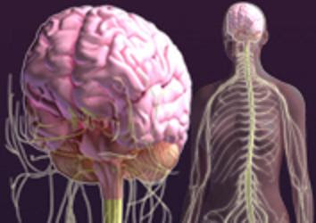
The Human Nervous System


Blue is PNS while red is CNS.


The peripheral nervous system (PNS)
resides or extends outside the central nervous system.

The central nervous system (CNS),
which consists of:
The brain
The spinal cord
To serve the limbs and organs. Unlike the central nervous system, however,

The PNS is not protected by bone, leaving it exposed to toxins and mechanical injuries.
The peripheral nervous system is divided into:
a. The somatic nervous system
b. The autonomic nervous system

the Central Nervous System

The central nervous system (CNS) of the vertebrate nervous system which is enclosed in meninges. It contains the majority of the nervous system, and consists of the brain (in vertebrates which have brains), and the spinal cord. Together with the peripheral nervous system, it has a fundamental role in the control of behavior. The CNS is contained within the dorsal cavity, with the brain within the cranial cavity, and the spinal cord in the spinal cavity. The brain is also protected by the skull, and the spinal cord is, in vertebrates, also protected by the vertebrae.

Nervous System Organization

The nervous system is composed of the central nervous system and the peripheral nervous system. The central nervous system which includes the brain and spinal cord, processes and coordinates all incoming sensory information and outgoing motor commands, and it is also the seat of complex brain functions such as memory, intelligence, learning, and emotion.
The peripheral nervous system includes all neural tissue outside of the central nervous system. It is responsible for providing sensory, or afferent, information to the central nervous system and carrying motor, or efferent, commands out to the body’s tissues. Voluntary motor commands, such as moving muscles to walk or talk, are controlled by the somatic nervous system, while involuntary motor commands, such as digestion and heart beat, are controlled by the autonomic nervous system. The autonomic nervous system is further divided into two systems. The sympathetic nervous system, sometimes called the “fight or flight” system, increases alertness, stimulates tissue, and prepares the body for quick responses to unusual situations. In contrast, the parasympathetic nervous system, sometimes called the “rest and repose” system, conserves energy and controls sedentary activities, such as digestion.
source: wiki/encarta encyclopedia

What is the Nervous System?

The nervous system
is a highly specialized network whose principal components are nerves called neurons. Neurons are interconnected to each other in complex arrangements and have the property of conducting, using electrochemical signals, and a great variety of stimuli both within the nervous tissue as well as from and towards most of the other tissues. Thus, neurons coordinate multiple functions in organisms. Nervous systems are found in many multicellular animals but differ greatly in complexity between species.
The nervous system has two divisions: the somatic, which allows voluntary control over skeletal muscle, and the autonomic, which is involuntary and controls cardiac and smooth muscle and glands. The autonomic nervous system has two divisions: the sympathetic and the parasympathetic. Many, but not all, of the muscles and glands that distribute nerve impulses to the larger interior organs possess a double nerve supply; in such cases the two divisions may exert opposing effects. Thus, the sympathetic system increases heartbeat, and the parasympathetic system decreases heartbeat. The two nervous systems are not always antagonistic, however. For example, both nerve supplies to the salivary glands excite the cells of secretion. Furthermore, a single division of the autonomic nervous system may both excite and inhibit a single effector, as in the sympathetic supply to the blood vessels of skeletal muscle. Finally, the sweat glands, the muscles that cause involuntary erection or bristling of the hair, the smooth muscle of the spleen, and the blood vessels of the skin and skeletal muscle are actuated only by the sympathetic division.
source: wiki/encarta encyclopedia

Sunday, July 6, 2008

EXPLORE THE GREATEST DISCOVERY

The medical discoveries of the past 1,000 years have saved countless lives and have doubled the life expectancy of people in many parts of the world. In this October 1999 Encarta Yearbook article, physicians Meyer Friedman and Gerald W. Friedland explore ten discoveries that fundamentally changed the way scientists and physicians were able to improve human health.
Microsoft ® Encarta ® 2008. © 1993-2007 Microsoft Corporation. All rights reserved.
The Greatest Medical Discoveries of the Millennium

By Meyer Friedman and Gerald W. Friedland
During the last 1,000 years, a series of monumental discoveries revolutionized the practice of medicine. These discoveries have saved millions of lives and brought about remarkable improvements in the health of entire populations.
One thousand years ago the average person in Europe could expect to live about 30 years. Of 100 children born alive, 40 would die before their first birthday. Disease and infections, largely the result of squalid living conditions, claimed untold numbers of lives. The average life span throughout the industrialized West has now more than doubled the average in 1000—to about 76.5 years for babies born in 1997 in the United States. Infant mortality rates are now a mere fraction of what they were 1,000 years ago.
Medical breakthroughs during the next millennium will probably bring even longer, healthier lives. Advances in genetics, for example, offer hope of new treatments to cure serious diseases such as cancer, eliminate genetic defects from families, and possibly even slow the aging process. And recent developments in transplantation technology suggest that soon it may be possible to grow replacement organs in the laboratory. But none of these anticipated breakthroughs would be possible—even thinkable—without the pioneering medical discoveries of the last 1,000 years.
Sorting through literally thousands of medical discoveries made during the past millennium to determine the ten most important is a challenge. How is the significance of a discovery measured? One factor is paramount: The most fundamental breakthroughs led to multiple other discoveries that eventually reshaped medicine and affected millions, even billions, of people. To be sure, changes in medical practices often lagged far behind the initial discovery; effective therapies sometimes emerged only after many years. These discoveries have attained the status of millennial importance not because they quickly helped physicians save lives, but because they fundamentally shifted the way scientists and physicians thought about human health. In so doing, the discoveries opened vast new fields of research that would revolutionize medicine and save the lives of incalculable numbers of people.
These seminal breakthroughs are, in the order of their discovery, human anatomy, circulation of blood, bacteria, vaccination, surgical anesthesia, X rays, blood typing, tissue culture, antibiotics, and the structure of deoxyribonucleic acid (DNA).
source: encarta encyclopedia


Setting the Stage

The practice of medicine is as old as civilization itself, but the Greeks are generally credited with inventing the science of medicine—using observation and experience rather than appeals to supernatural forces to treat disease. Although Greek medical knowledge was passed on to the conquering Romans, it fell into obscurity as the Roman Empire collapsed in the early Middle Ages.
A period of stagnation in the sciences, combined with sporadic epidemics of the bubonic plague, smallpox, and other diseases, reinforced the turn toward superstition and magical treatments in medieval Europe. Only fragments of the ancient medical learning survived. Many people viewed disease as a form of punishment for sins or as the result of demonic forces. Prayer was a standard form of treatment.
Western medicine received a major boost when the Italian universities of Salerno, Bologna, and Padua established medical faculties in the 9th and 10th centuries. By the 12th century, the University of Paris in France and Oxford University in England had also founded faculties of medicine. These institutions provided facilities for research, set examination requirements for graduating physicians, and laid the foundations for the extraordinary revival of Western medicine in the 16th and 17th centuries—a revival that has continued to this day.

source: encarta encyclopedia
Human Anatomy

Before modern medical science could emerge, medical practitioners needed an accurate understanding of human anatomy. Without clear descriptions of the structure of the human body, it was impossible to learn what different bodily parts actually do. Once researchers understood how parts of the body worked, they were better able to devise medical therapies to restore proper functions.
Amazingly, no one knew much about human anatomy until 1543, when the Belgian anatomist Andreas Vesalius wrote De Humani Corporis Fabrica, Libri Septem (On the Structure of the Human Body, in Seven Books). For centuries, exploration of the anatomy of human corpses was forbidden. In medieval Europe, knowledge about anatomy was based largely on the teachings of the Roman physician Galen (129-199?). Galen's anatomical descriptions were based on dissections of animals, which differ in many ways from humans. But contradicting Galen was dangerous because the powerful Roman Catholic Church accepted his findings as gospel. A few brave souls had tried to correct some of Galen's errors, but their work was lost for centuries.
Ambitious, driven, and ruthless, 23-year-old Vesalius received his medical degree in 1537 from the University of Padua and was immediately appointed head of surgery and anatomy there. As a student, and later as a scientist, he recovered human corpses from cemeteries late at night. He even encouraged his students to note patients who were at death's door so that he could steal their bodies for dissection before they were buried. Vesalius slept, night after night, with corpses in his own bedroom, and he hired Italy's greatest artists to draw what he found.
In 1543 Vesalius completed his seven-book masterpiece, richly illustrated with more than 200 magnificent drawings. Many consider it one of the greatest medical books ever published. This monumental work gave medicine a precious gift: For the first time, human anatomy was based on careful dissection and observation rather than on a rigid orthodoxy rooted in ancient texts
.
source: encarta encyclopedia
Circulation of Blood

English physician William Harvey's discovery of what the heart does and how the blood circulates is widely regarded as the single greatest medical achievement of all time: It established the principle of doing experiments in medicine to learn how the body's organs and tissues function. Published in 1628, Harvey's groundbreaking book Exercitatio Anatomica de Motu Cordis et Sanguinis in Animalibus (Anatomical Essay on the Motion of the Heart and Blood in Animals) spurred research into the mechanical functions of many bodily processes, including respiration, digestion, metabolism, and reproduction.
Harvey received his medical degree from the University of Padua, where he learned one very important fact: Veins have valves that permit blood to travel in only one direction. However, the exact role of the valves was unclear.
Realizing that it was still dangerous to contradict Galen, who had claimed that the liver not only makes the body's blood but also pumps it through the body, Harvey decided to study blood flow by operating on live animals. For a period of 12 years Harvey conducted his experiments before members of the influential Royal College of Physicians in London, England. He wanted their support for his book, which praised Galen while challenging many of his ideas.
In the 8th chapter of his 17-chapter book, Harvey carefully introduced the revolutionary idea that blood goes in a circle in the body, traveling from the heart to the arteries to the veins and back to the heart. The next 9 chapters proved, in wonderfully clear English, that he was right.
In a series of brilliant experiments in animals and humans, Harvey demonstrated how blood circulates in the body. When an artery was blocked, the veins draining this artery collapsed. When a vein was blocked, it swelled below the blockage and collapsed above it, but the swelling disappeared when the blockage was removed. He also showed that the valves in veins allow blood to flow only in the direction of the heart. Together, these discoveries proved that blood moves in a circle in the body—that is, there is a “circulation.”
source: encarta encyclopedia
Bacteria

After the momentous medical breakthroughs of Vesalius and Harvey came the 17th-century discovery of one of the human body's greatest enemies: bacteria. This discovery eventually led to the realization that exposure to certain microorganisms could cause disease. It also prompted new theories of antiseptics that sharply lowered mortality rates from surgery.
Antoni van Leeuwenhoek, a part-time janitor and haberdasher working in Delft, Holland, discovered bacteria and other microorganisms using a microscope that he built himself. Through the influence of a friend, a Dutch physician, he was invited to write letters to the Royal Society of London—a group dedicated to the advancement of science. These letters were translated from Dutch into English and published in the society's journal Philosophical Transactions.
Leeuwenhoek's most famous letter was published on March 16, 1677. In this letter he described looking at a drop of rainwater through his microscope. The drop was taken from a tub where it had been allowed to stand for several days. To his amazement, he saw exceedingly tiny animals, known today as protozoa, swimming in the water. He also observed other equally small animals that did not move at all, now known as bacteria. No one at the Royal Society knew anything about these little creatures, which Leeuwenhoek called animalcules. At the request of the stunned members of the Royal Society, several of the most respected citizens of Delft were asked to verify Leeuwenhoek's microscopic findings. They did so, and in 1680 Leeuwenhoek was elected a fellow of the prestigious Royal Society.
Later discoveries extended the significance of Leeuwenhoek's work, especially the superb finding by the German scientist Robert Koch in 1876. Koch found that the microscopic anthrax bacillus could actually cause a fatal human disease. Until Koch's discovery, many scientists thought it absurd that microscopic creatures could harm much larger animals, such as humans. In 1882 Koch showed that another kind of bacterium, the tubercle bacillus, caused tuberculosis, a discovery for which he won the Nobel Prize in 1905.
Unlike Koch, who was a country physician when he made his epochal discoveries, the French chemist and biologist Louis Pasteur disliked physicians so much that he would not have them as workers in his laboratory. Despite his disdain for physicians, he was deeply fascinated by diseases of various kinds. Pasteur discovered that putrefaction (a decomposition of organic substances) is caused by microorganisms that float in the air. Pasteur learned that he could prevent putrefaction by subjecting organic substances to moderate but not extreme heat, a process known as pasteurization.
In 1865 Joseph Lister, an English surgeon, read of Pasteur's research on putrefaction. Lister recalled that whereas simple bone fractures invariably healed, compound fractures (bone fractures bursting the skin) almost always began to putrefy. Lister was certain that this dangerous, infectious process was caused by the wretched microorganisms that Pasteur had described. To test his theory, Lister covered his patients' compound fractures, previously exposed to the air, with linen strips soaked in carbolic acid. He believed the carbolic acid might exterminate the airborne microorganisms.
Lister treated compound fractures and open surgical wounds with carbolic acid for nine months. During this time, he did not observe a single infection in his surgical ward. The results of his experiments, published in 1867, gave rise to antiseptic surgery. Although Lister's antiseptic technique initially encountered resistance from other physicians, it soon became widely accepted, and deaths due to infections in the operating room plummeted.

source: encarta enyclopedia
Vaccination

Smallpox, once a common viral infection that claimed millions of lives in periodic epidemics around the globe, is now considered fully eradicated. Much of the credit for this medical triumph belongs to the English physician Edward Jenner, who in 1796 developed the first effective vaccine against smallpox. Jenner's discovery laid the foundations for the science of immunology. Vaccines are now used to control and prevent diphtheria, hepatitis, influenza, meningitis, polio, tetanus, typhoid fever, whooping cough, and many other diseases that once plagued humankind.
In Jenner's day a procedure called variolation was used to protect people against smallpox. The procedure involved scratching a bit of the substance from smallpox blisters—obtained from a person with a mild case of the disease—into a healthy person's arm. Hopefully, a mild case of the disease would develop and pass, but the procedure was often deadly.
Jenner, orphaned at the age of five, was born and raised in the tiny English village of Berkeley, near Bristol. At the age of 13, Jenner was apprenticed to a country surgeon. Shortly after, milkmaids told him that after they contracted cowpox, a harmless disease confined usually to their hands and arms, they never got smallpox.
Following his training with the famous surgeon John Hunter in London, Jenner returned to Berkeley and devised an experiment to learn whether cowpox could protect against smallpox. On May 14, 1796, Jenner made two small scratches on the arm of an eight-year-old boy named James Phipps. On those scratches he rubbed fluid from a milkmaid's cowpox blister. Eight days later, Phipps developed small cowpox blisters on the scratches. On July 1 Jenner variolated Phipps with fluid from a smallpox blister. Phipps never got even a mild case of smallpox.
Jenner had made two important discoveries: Cowpox protects against smallpox, and cowpox could be transmitted from person to person. He subsequently vaccinated another eight children, including his own son, experimenting further with his new technique. In 1798 Jenner submitted his findings to Philosophical Transactions, but his work was rejected. After further experiments, he published his results himself, paying for the printing.
Vaccination was initially viewed as unnatural, and the technique encountered significant opposition for decades. More than 80 years passed before Pasteur, drawing on Jenner's work, opened the way for the development of modern preventive vaccines. In the end, however, Jenner received an honorary degree from Oxford University for his groundbreaking work.
source: encarta encyclopedia
Surgical Anesthesia

Until the discovery of anesthesia by Crawford Williamson Long in 1842, surgery was an excruciating ordeal, usually attempted only in cases of dire injury or illness. Some patients used alcohol or opium to lessen the pain; others recited verse. Passing out was a blessing. Surgeons worked at breakneck speed to get in and out of the patient's body as quickly as possible. Anesthesia changed all this, permitting surgeons to work at a slower, more careful pace.
The Spaniard Lullius discovered ether, an organic solvent, in 1275, but its anesthetic properties were unknown. In the early 1800s people inhaled ether at parties to make themselves high. Long, a physician in Jefferson, Georgia, frequently prepared ether at the request of his friends. One evening he used it himself at a so-called ether frolic and badly bruised himself while he was high. Yet, Long noticed, he felt no pain.
On March 30, 1842, Long convinced James Venable, who had two cysts in his neck and was terrified at the prospect of surgery, to try ether. Venable did, and the ether made him unconscious. The operation was a success, and Venable, amazed, felt no pain at all. During the next four years, Long successfully used ether as anesthesia on eight patients.
But in 1842 the physician Charles Jackson and a dentist, William Morton, learned what Long was doing with ether and stole his secret, possibly after visiting Jefferson. Morton used ether to anesthetize two patients at Boston's Massachusetts General Hospital on October 16, 1846, in front of an audience of famous surgeons. The results were published, and anesthesia was soon used around the world. Long received no credit for the discovery.
The claims of Jackson, Morton, Long, and others created bitter quarrels about who actually invented surgical anesthesia. The Congress of the United States even took up the matter, debating the issue for 16 years without ever deciding who first introduced ether as an anesthetic procedure. But the use of anesthesia developed rapidly. Later, scientists found new anesthetic agents, developed better methods of administering anesthetic gases, and eventually discovered the use of local anesthesia.

source: encarta encyclopedia
X rays

The development of X-ray photography, or radiology, was an enormous leap into the future: For the first time, physicians could see inside the body without opening it. With X rays surgeons could quickly diagnose fractures, tumors, and other ailments and plan more intricate operations. As a result, surgery rapidly grew in sophistication.
Early in 1895 German physicist Wilhelm Conrad Roentgen was experimenting with a Crookes tube—a pear-shaped glass tube emptied of air with electrodes (metal wires) sealed into opposite ends of the tube. When the negative electrode, or cathode, received a high-voltage electric current, it glowed white-hot and emitted a stream of invisible, electrically charged particles called cathode rays. These rays moved towards the positive electrode, or anode, of the tube. If only a little air remained inside the Crookes tube, the cathode rays striking the glass at the other end of the tube would produce a yellow-green fluorescence.
Roentgen's experiments had confirmed another physicist's observation that cathode rays could pass through an aluminum-covered window in the wall of a Crookes tube. To discover whether cathode rays could also go through the glass wall of a Crookes tube, he placed a piece of paper coated with a barium salt near the tube's anode. Such paper was known to fluoresce when hit with cathode rays. He expected the fluorescence to be faint, so he covered the tube with black cardboard to block the tube's fluorescence and to better help him see. He also darkened his laboratory.
While testing the tube to see whether any fluorescence was visible through the cardboard, Roentgen noticed a strange glow some distance away. Lighting a match for light to see, he discovered that the glow was coming from another piece of coated paper that was about a meter away from the Crookes tube. Repeatedly turning his tube on and off, he learned that the paper only glowed when the tube was on. The paper still fluoresced when he moved it even farther away and even when he shielded it, first with a pack of cards and then with a book.
Roentgen knew that cathode rays were not strong enough to cause this distant fluorescence. There could only be one explanation: The Crookes tube was producing previously unknown kinds of electromagnetic waves, which he later called X rays. Further experiments showed that these new X rays would not go through lead and would go only partly through other metals.
In December 1895 he X-rayed his own fingers holding a small lead pipe. To his astonishment, the developed pictures revealed not only the shadow of the pipe but also the bones of two of his fingers. He later X-rayed the left hand of his wife, Bertha, who had two gold rings on her fourth finger, and showed the terrified woman a picture of her own bones, complete with rings.
Roentgen's preliminary report, entitled On a New Kind of X Ray, was published only a few days after he submitted it, a record for rapid publication in science. In 1901 Roentgen became the first scientist ever to receive the Nobel Prize in physics.
X rays were used immediately for medical diagnosis but did not become a routine procedure until the 1920s. In the decades that followed, technological advances allowed X rays to outline individual organs and organ systems, as well as arteries and veins. In 1972 researchers developed the computerized tomography (CAT) scan, a sophisticated X-ray technology that produces computer-generated cross-sectional views of the body.
source: encarta encyclopedia
Blood Typing

At the dawn of the 20th century, the Austrian physician Karl Landsteiner made the extraordinary discovery that human blood could be grouped into several different types. This discovery made possible the transfer of blood from one human to another—a medical breakthrough that has saved countless lives.
Prior to Landsteiner's pioneering work, there were few reports on the transfer of blood from one human to another. In 1668 Jean Baptiste Denis, the French physician to King Louis XIV, dared to transfuse a man with sheep's blood. The man eventually died and Denis was arrested for murder. Transfusions were quickly banned in France and England. Other attempted transfusions using human blood were frequently unsuccessful, and patients often died due to blood incompatibility.
In 1900 Landsteiner made the brilliant observation that human blood contains what he called isoagglutinins. These proteins are capable of agglutinating, or clotting, the red blood cells of blood samples containing isoagglutinins different from their own. He thus was able to divide blood into three types: A, B, and O. A rare fourth type, AB, was later discovered.
Landsteiner showed that the sera of two blood samples containing the same isoagglutinins would not clot the red cells of either blood. This discovery permitted the development of a system for safe blood transfusions. For this gift to humankind, Landsteiner received the Nobel Prize in physiology or medicine in 1930.
source: encarta encyclopedia
Tissue Culture

In 1907 American biologist Ross Granville Harrison stumbled upon the amazing discovery that living tissues could be cultured, or grown, outside the body. Although Harrison could not have known it at the time, his discovery would become one of the most valued techniques in medicine. Tissue culture has opened up new ways to study the development of genes (the basic units of heredity), embryos, tumors, toxins, and the pathogens that cause numerous diseases. The technique is also used to produce medicines, vaccines, and replacement tissues, as well as to clone animals, such as Dolly, the famous sheep.
In late summer of 1906 Harrison, an expert on embryos, wanted to solve what was then an important problem in biology. Harrison set out to determine whether nerve fibers grow in local tissues of the body or originate in nerve cells of the brain. He had to devise a new way to study the problem because all the available living specimens contained both nerves and surrounding tissue.
Harrison decided to study living nerves in the absence of any surrounding tissue. To do this, he isolated a portion of the hindbrain of a tiny living frog embryo. To keep his specimen alive, he immersed it in fresh frog lymph (a diluted blood plasma carried by the lymphatic system) and placed it under a cover slip so he could examine it with a microscope. The frog lymph quickly clotted, like blood, and he sealed it with wax to prevent evaporation or contamination of the specimen.
Using a microscope, Harrison discovered that the nerve fiber did actually come from the brain, not the surrounding tissue. Harrison noticed something else: The frog's brain cells were still growing, even though they were no longer in the frog's body. Harrison found the answer to the question he was asking and, at the same time, invented what would become the science of tissue culture. Harrison reported his result in May 1907. Since then tissue culture has allowed researchers to learn more about the basic mechanisms of disease than had been learned in the previous 500 years.

source: encarta encyclopedia
Antibiotics

The discovery of antibiotics opened a whole new front in the war against disease. Armed with antibiotics, which act by killing bacteria or inhibiting their growth, scientists have mounted a major assault on cholera, pneumonia, tetanus, tuberculosis, and many other deadly bacterial infections that had previously struck people down relentlessly.
Some of the most important breakthroughs in science occur unexpectedly, and the discovery of penicillin—perhaps the world's most widely used antibiotic—is one such example. The British bacteriologist Sir Alexander Fleming is credited with discovering penicillin, although other scientists before him had noticed that the mold Penicillium notatum prevented the growth of some types of bacteria. So Fleming's discovery was actually a rediscovery.
In September 1928 Fleming was preparing to take a short vacation with his family, when a series of almost unbelievably lucky events occurred. Just before leaving, Fleming decided to cultivate staphylococci to study when he returned. This was the first piece of luck. He could have picked any bacterium to study, but he happened to pick one that would turn out to be susceptible to penicillin.
Fleming opened a petri dish for a few seconds to put the staphylococci inside. Ordinarily, no mold spores would have a chance to get in the dish, but two floors below his laboratory, another scientist was studying the mold Penicillium notatum. Millions of very light mold spores floated in the air, up the staircase and the elevator shaft, through the always-open doors of Fleming's laboratory, and into the open dish where, luckily, he was just putting the staphylococci.
Fleming, preoccupied with his vacation, left the petri dish on the laboratory bench instead of putting it in a warm incubator. This was lucky, too, because the bacteria and Penicillium notatum usually grow at different temperatures. Staphylococci multiply at relatively high temperatures, while Penicillium multiplies at lower temperatures. While Fleming was away the temperature turned out to be perfect for Penicillium, but not so good for the staphylococci, which grew slowly. The Penicillium mold thrived and secreted penicillin, which oozed around the dish, preventing the growth of staphylococci and leaving the Penicillium mold isolated from small bacterial colonies in the dish.
Fleming, upon his return, immediately realized what had happened, and he conducted other tests to learn what other bacteria this mysterious mold stuff could kill. He also tried to make pure penicillin, but did not succeed. Fleming believed that the mold substance, which he named penicillin, could be rubbed onto a cut or a scrape to prevent an infection. A few years later, however, Fleming gave up studying the mold.
As a consequence, penicillin was nearly forgotten until the beginning of World War II (1939-1945). Scientists at Oxford University in England showed that penicillin could prevent bacterial infections in animals and humans, and they devised a technique to mass-produce pure penicillin. The scientists encouraged companies in the United States to manufacture penicillin in vast quantities, and the new drug was credited with saving thousands of lives during the war. In 1945 Fleming and two of the Oxford scientists, Sir Howard Florey and Ernst B. Chain, received the Nobel Prize in physiology or medicine.
source: encarta encyclopedia
DNA

Perhaps the greatest medical breakthrough of the 20th century is the discovery of the structure of deoxyribonucleic acid (DNA—the molecular basis of heredity). Knowledge of DNA's chemical structure allowed scientists to understand for the first time how DNA replicates itself and passes information from one generation to the next. This monumental discovery has already revolutionized many aspects of medicine, permitting the development of a vast range of genetically engineered drugs, hormones, and other useful substances. Even more radical changes are afoot. In the new millennium, scientists are expected to have access to a complete map of the human genetic code, which should help them trace the genetic causes of all inherited diseases and search out possible cures.
The Swiss physician Friedrich Miescher isolated DNA for the first time in 1869, but the function of the chemical, which is found only in the nucleus of cells, was unknown. As the years passed, scientists learned that DNA contained phosphate, a sugar called deoxyribose, and four different compounds called nucleotide bases.
In 1944 the Canadian-born American physician and bacteriologist Oswald T. Avery and his colleagues showed, in a series of experiments on bacteria, that DNA transmitted genetic information. Prior to Avery's groundbreaking work, many biochemists believed that proteins were the source of genetic information.
By 1950 two groups of scientists were in hot pursuit of the structure of DNA. One of the groups was at Cavendish Laboratory in Cambridge, England. The other group, at King's College, London, consisted of Maurice Wilkins, a physicist, and Raymond Gosling, a graduate student. They were joined in 1951 by Rosalind Franklin, an expert in X-ray crystallography (a technique that uses a tiny beam of X rays to create images of the structural relationships between atoms and molecules of chemical substances). Photographs of images produced by X-ray crystallography are called X-ray diffraction photographs.
In 1950 Wilkins received a uniquely pure sample of DNA from a Swiss physicist. From this sample, he was able to pick out single DNA fibers with a glass rod. Wilkins and Gosling X-rayed these fibers in 1950, as did Franklin when she joined the laboratory in 1951.
However, a misunderstanding caused Wilkins and Franklin each to think they were in charge of X-ray crystallography, and they did not cooperate. When Franklin left the team in 1952, she was ordered to submit all of the X-ray diffraction photographs to Wilkins. One of these photographs showed that the DNA molecule had the shape of a double helix, a structure resembling a twisted ladder.
In the meantime the American biologist James Watson attended a meeting in Naples, Italy, in 1950, in which he saw one of Wilkins's X-ray diffraction photographs. Watson immediately thought the molecule might be a double helix. In the fall of 1951 he joined the team of scientists at Cavendish Laboratory, where he convinced a British biophysicist, Francis Crick, that a combination of model building—using plastic balls, wires and steel plates—and X-ray crystallography could lead them to the structure of DNA.
The double helix by itself, however, was not the only secret to the DNA molecule. Its entire chemistry needed to be explained. Watson, unbeknownst to Wilkins, was now convinced that DNA had a helical structure and was working feverishly with Crick on their increasingly complex model of the molecule, which they finished during the second week of 1953. This model incorporated all the known chemical components of DNA and closely matched the diffraction pattern observed in Wilkin's photograph. Watson and Crick accurately deduced that the two strands of the double helix separated before cellular division, providing templates, or patterns, for the creation of two new DNA molecules identical to the original.
Watson and Crick sent Wilkins a copy of their manuscript, which took advantage, of course, of what Wilkins and Franklin had done, and which Wilkins thought was his own. After reading their manuscript, Wilkins sent them a letter that began, “I think you are a couple of old rogues.…”
On April 25, 1953, the journal Nature published one article from the Cambridge laboratory and two from King's College in London on the molecular structure of DNA. Many felt that the key to life itself had been revealed. Wilkins, Watson, and Crick shared the 1962 Nobel Prize in physiology or medicine.
source: encarta encyclopedia
Looking Ahead

The discovery of the structure of DNA—like the discovery of X rays at the end of the 19th century or the detection of bacteria more than two centuries before that—has radically altered medicine and opened previously unknown frontiers. Equipped with a map of the human genome (the complete genetic code), researchers in the next millennium hope to root out the genetic causes of a wide range of inherited diseases, from schizophrenia to cystic fibrosis to hemophilia to many types of cancer. Of perhaps greater significance, many scientists believe that advances in molecular genetics are setting the course for fundamental changes in the diagnosis and treatment of disease. Instead of merely treating the symptoms of disease, as they do now, physicians of the next millennium may develop the ability to routinely identify and correct the causes of disease before symptoms appear.
And yet, despite Western medicine's stunning success in fighting disease and extending human life, the health of much of the developing world is worsening. Modern vaccines and antibiotics would save tens of millions of lives each year throughout the developing world, where people are continuously struck down by malaria, tuberculosis, polio, pneumonia, and other easily treatable disorders. And the vast majority of people with acquired immune deficiency syndrome (AIDS) now live in the developing world, where access to costly life-extending medications is beyond the reach of most of those infected. Finding ways to extend the magnificent contributions of Western medicine to those who need it most therefore constitutes one of the greatest medical challenges of the coming millennium.

source: encarta encyclopedia

The Authors

Meyer Friedman is a founder of the San Francisco-based Meyer Friedman Institute, a nonprofit research group. Gerald W. Friedland is a professor emeritus of radiology at Stanford University. Friedman and Friedland are coauthors of Medicine's 10 Greatest Discoveries, recently published by Yale University Press.

For further reading:
Fisher, Richard B. Edward Jenner, 1749-1823. Andre Deutsch, 1991.
Fradin, Dennis Brindell. “We Have Conquered Pain:” The Discovery of Anesthesia. Margaret McElderry, 1996.
Friedman, Meyer, and Gerald W. Friedland. Medicine's 10 Greatest Discoveries. Yale University Press, 1998.
Judson, Horace Freeland. The Eighth Day of Creation: Makers of the Revolution in Biology. Simon and Schuster, 1996.
Kevles, Bettyann Holtzmann. Naked to the Bone: Medical Imaging in the Twentieth Century. Rutgers University Press, 1996.
O'Malley, C. D. Andreas Vesalius of Brussels, 1514-1564. University of California Press, 1964.
Porter, Roy. The Cambridge Illustrated History of Medicine. Cambridge University Press, 1996.
source: encarta encyclopedia

Looking ahead on INSIGHT

From the Notebooks of
Leonardo da Vinci

The life and structure of things

Although the painter's eye sees but the surface of things it must in rendering the surface discern and interpret the organic structure that lies beneath. The representation of the human figure entails a knowledge of its anatomy and its proportions. Landscapes entail a study of the morphology of plants, of the formation of the earth, of the movements of water and wind.
This work must begin with the conception of man, and describe the nature of the womb and how the foetus lives in it, up to what stage it resides there and in what ways it quickens into life and feeds. Also its growth and what interval there is between one stage of growth and another. What it is that forces it out from the body of the mother, and for what reasons it sometimes comes out of the mother's womb before due time.
Then I will describe which are the members which after the boy is born, grow more than the others, and determine the proportions of a boy of one year. Then describe the fully grown man and woman, with their proportions, and the nature of their complexions, colour, and physiognomy.
Then how they are composed of veins, tendons, muscles, and bones. Then in four drawings represent four universal conditions of men. That is, mirth with various acts of laughter; and describe the cause of laughter. Weeping in various aspects with its causes. Strife with various acts of killing: flight, fear, ferocity, boldness, murder, and everything pertaining to such conditions. Then represent Labour with pulling, thrusting, carrying, stopping, supporting, and such-like things.
Further, I would describe attitudes and movements. Then perspective concerning the function of the eye; and of hearing—here I will speak of music—and treat of the other senses—and describe the nature of the five senses. This mechanism of man we will demonstrate with [drawings of] figures.
source: encarta encyclopedia
(a) Proportion

The theory of proportions had a great fascination for Renaissance artists. Their canons were not only intended as a means of artistic workmanship, they were meant to achieve harmony. Proportions in painting, sculpture, and architecture were like harmony in music and gave intense delight.
Proportion is not only found in numbers and measurements but also in sounds, weights, time, and position, and whatever power there may be.
The Roman architect Vitruvius had transmitted some data of a Greek canon for the proportions of the human figure and these were revived in Renaissance time. A drawing by Leonardo, now in the Academy at Venice, was reproduced in an edition of Vitruvius' book, published in 1511, in order to illustrate the statement that a well-made human body with arms outstretched and feet together can be inscribed in a square; while the same body spread-eagled occupies a circle described around the navel. The proportions of the human body are here related to the most perfect geometric figures and may be said to be integrated into the spherical cosmos. Leonardo endeavoured to verify and elaborate Vitruvius' mathematical formulae in order to put them on a scientific basis by empirical observations, and for this purpose he collected data from living models.
Geometry is infinite because every continuous quantity is divisible to infinity in one direction or the other. But the discontinuous quantity commences in unity and increases to infinity, and as it has been said the continuous quantity increases to infinity and decreases to infinity. And if you give me a line of twenty braccia I will tell you how to make one of twenty-one.
Every part of the whole must be in proportion to the whole … I would have the same thing understood as applying to all animals and plants.
From painting which serves the eye, the noblest sense, arises harmony of proportions; just as many different voices joined together and singing simultaneously produce a harmonious proportion which gives such satisfaction to the sense of hearing that listeners remain spellbound with admiration as if half alive. But the effect of the beautiful proportion of an angelic face in painting is much greater, for these proportions produce a harmonious concord which reaches the eye simultaneously, just as a chord in music affects the ear; and if this beautiful harmony be shown to the lover of her whose beauty is portrayed, he will without doubt remain spellbound in admiration and in a joy without parallel and superior to all other sensations.
The painter in his harmonious proportions makes the component parts react simultaneously so that they can be seen at one and the same time both together and separately; together, by viewing the design of the composition as a whole; and separately by viewing the design of its component parts.
Vitruvius, the architect, says in his work on architecture that the measurements of the human body are distributed by nature as follows: 4 fingers make 1 palm; 4 palms make 1 foot; 6 palms make 1 cubit; 4 cubits make a man's height; and 4 cubits make one pace; and 24 palms make a man; and these measures he used in buildings.
If you open your legs so much as to decrease your height by 1/14 and spread and raise your arms so that your middle fingers are on a level with the top of your head, you must know that the navel will be the centre of a circle of which the outspread limbs touch the circumference; and the space between the legs will form an equilateral triangle.
The span of a man's outspread arms is equal to his height.
From the roots of the hair to the bottom of the chin is the tenth part of a man's height; from the bottom of the chin to the crown of the head is the eighth of the man's height; from the top of the breast to the crown of the head is the sixth of the man; from the top of the breast to the roots of the hair is the seventh part of the whole height; from the nipples to the crown of the head is a fourth part of the man. The maximum width of the shoulders is the fourth part of the height; from the elbow to the tip of the middle finger is the fifth part; from the elbow to the end of the shoulder is the eighth part. The complete hand is the tenth part. The penis begins at the centre of the man. The foot is the seventh part of the man. From the sole of the foot to just below the knee is the fourth part of the man. From below the knee to where the penis begins is the fourth part of the man.
The distance between the chin and the nose and that between the eyebrows and the beginning of the hair is equal to the height of the ear and is a third of the face.
The length of the foot from the end of the toes to the heel goes twice into that from the heel to the knee, that is, where the leg-bone joins the thigh bone. The hand to the wrist goes four times into the distance from the tip of the longest finger to the shoulder-joint.
A man's width across the hips is equal to the distance from the top of the hip to the bottom of the buttock, when he stands equally balanced on both feet; and there is the same distance from the top of the hip to the armpit. The waist, or narrower part above the hips, will be half-way between the armpits and the bottom of the buttock.
Every man at three years is half the full height he will grow to at last.
There is a great difference in the length between the joints in men and boys. In man the distance from the shoulder joint to the elbow, and from the elbow to the tip of the thumb, and from one shoulder to the other, is in each instance two heads, while in a boy it is only one head; because Nature forms for us the size which is the home of the intellect before forming what contains the vital elements.
Remember to be very careful in giving your figures limbs that they should appear to be in proportion to the size of the body and agree with the age. Thus a youth has limbs that are not very muscular nor strongly veined, and the surface is delicate and round and tender in colour. In man the limbs are sinewy and muscular; while in old men the surface is wrinkled, rugged and knotty, and the veins very prominent.
Microsoft ® Encarta ® 2008. © 1993-2007 Microsoft Corporation. All rights reserved.
(b) The Anatomy and Movement of the Body

The human body is a complex unity within the larger field of nature, a microcosm wherein the Elements and Powers of the universe were incorporated. In order to study its structure Leonardo dissected corpses and examined bones, joints, and muscles separately and in relation to one another, making drawings from many points of view and taking recourse to visual demonstration since an adequate description could not be given in words. According to him such visual demonstrations gave 'complete and accurate conceptions of the various shapes such as neither ancient nor modern writers have ever been able to give without an infinitely tedious and confused prolixity of writing and of time.' Moreover, there are not only the various points of view, the infinity of aspects to be considered, there are also the continuous successions of phases in movements. The circular movements of shoulder, arm, and hand, for instance, is suggestive of a pictorial continuity such as we may see on a strip of film.
The study of structure included that of function, of the manner in which actions and gestures were performed, how the various muscles work together in bending and straightening the joints; how the weight of a body is supported and balanced. Leonardo looked upon anatomy with the eye of a mechanician. Each limb, each organ was believed to be designed and perfectly adapted to perform its special function. Thus the muscles of the tongue were made to produce innumerable sounds within the mouth enabling man to pronounce many languages. In his time divisions between the various branches of anatomy did not exist. He investigated problems of physiology and embryology, the systems of nerves and arteries. He anticipated the principle of blood circulation and prepared the ground for further analyses on many subjects.
You who say that it is better to watch an anatomical demonstration than to see these drawings, you would be right if it were possible to observe all the details shown in such drawings in a single figure, in which with all your cleverness you will not see or acquire knowledge of more than some few veins, while in order to obtain a true and complete knowledge of these, I have dissected more than ten human bodies, destroying all the various members and removing the minutest particles of the flesh which surrounded these veins, without causing any effusion of blood other than the imperceptible bleeding of the capillary veins. And as one single body did not suffice for so long a time, it was necessary to proceed by stages with so many bodies as would render my knowledge complete; this I repeated twice in order to discover the differences. And though you should have a love for such things you may perhaps be deterred by natural repugnance, and if this does not prevent you, you may perhaps be deterred by fear of passing the night hours in the company of these corpses, quartered and flayed and horrible to behold; and if this does not deter you, then perhaps you may lack the skill in drawing, essential for such representation; and if you had the skill in drawing, it may not be combined with a knowledge of perspective; and if it is so combined you may not understand the methods of geometrical demonstration and the method of estimating the forces and strength of muscles; or perhaps you may be wanting in patience so that you will not be diligent.
Concerning which things, whether or no they have all been found in me, the hundred and twenty books which I have composed will give verdict 'yes' or 'no.' In these I have not been hindered either by avarice or negligence, but only by want of time. Farewell.
source: encarta encyclopedia
How it is necessary for the painter to know the inner structure of man

The painter who has a knowledge of the nature of the sinews, muscles, and tendons will know very well in the movement of a limb how many and which of the sinews are the cause of it, and which muscle by swelling is the cause of the contraction of that sinew; and which sinews expanded into most delicate cartilage surround and support the said muscle.
Thus he will in divers ways and universally indicate the various muscles by means of the different attitudes of his figures; and will not do like many who, in a variety of movements, still display the same things in the arms, the backs, the breasts, and legs. And these things are not to be regarded as minor faults.
In fifteen entire figures there shall be revealed to you the microcosm on the same plan as before me was adopted by Ptolemy in his cosmography; and I shall divide them into limbs as he divided the macrocosm into provinces; and I shall then define the functions of the parts in every direction, placing before your eyes the representation of the whole figure of man and his capacity of movements by means of his parts. And would that it might please our Creator that I were able to reveal the nature of man and his customs even as I describe his figure.
Remember, in order to make sure of the origin of each muscle to pull the tendon produced by this muscle in such a way as to see this muscle move, and its attachment to the ligaments of the bones. You will make nothing but confusion in demonstrating the muscles and their positions, origins and ends, unless you first make a demonstration of thin muscles after the manner of threads; and in this way you will be able to represent them one over the other as nature has placed them; and thus you can name them according to the limb they serve, for instance the mover of the tip of the big toe, and of its middle bone or of the first bone, etc. And when you have given this information you will draw by the side of it the true form and size and position of each muscle; but remember to make the threads which denote the muscles in the same positions as the central line of each muscle; and so these threads will demonstrate the shape of the leg and their distance in a plain and clear manner.
source: encarta encyclopedia
Of the hand from within

When you begin the hand from within first separate all the bones a little from each other so that you may be able quickly to recognize the true shape of each bone from the palm side of the hand and also the real number and position in each finger; and have some sawn through lengthwise, so as to show which is hollow and which is full. And having done this replace the bones together at their true contacts and represent the whole hand from within wide open. The next demonstration should be of the muscles around the wrist and the rest of the hand. The fifth shall represent the tendons which move the first joints of the fingers. The sixth the tendons which move the second joints of the fingers. The seventh those which move the third joints of these fingers. The eighth shall represent the nerves which give them the sense of touch. The ninth the veins and the arteries. The tenth shall show the whole hand complete with its skin and its measurements; and measurements should also be taken of the bones. And whatever you do for this side of the hand you should also do for the other three sides—that is for the palmar side, for the dorsal side, and for the sides of the extensor and flexor muscles. And thus in the chapter on the hand you will give forty demonstrations; and you should do the same with each limb. And in this way you will attain thorough knowledge. You should afterwards make a discourse concerning the hands of each of the animals, in order to show in what way they vary. In the bear for instance the ligaments of the tendons of the toes are attached above the ankle of the foot.
Weight, force, and the motion of bodies and percussion are the four elemental powers in which all the visible actions of mortals have their being and their end.
After the demonstration of all the parts of the limbs or men and of the other animals you will represent the proper way of action of these limbs, that is in rising from lying down, in moving, running, and jumping in various attitudes, in lifting and carrying heavy weights, in throwing things to a distance, and in swimming; and in every action you will show which limbs and which muscles perform it, and deal especially with the play of the arms.
As regards the disposition of the limbs in movement you will have to consider that when you wish to represent a man who for some reason has to turn backwards or to one side you must not make him move his feet and all his limbs towards the side to which he turns his head. Rather must you make the action proceed by degrees and through the different joints, that is those of the foot, the knee, the hips, and the neck. If you set him on the right leg, you must make his left knee bend inwards and his left foot slightly raised on the outside; and let the left shoulder be somewhat lower than the right; and the nape of the neck is in a line directly over the outer ankle of the left foot. And the left shoulder will be in a perpendicular line above the toes of the right foot. And always set your figures so that the side to which the head turns is not the side to which the breast faces, since nature for our convenience has made us with a neck which bends with ease in many directions as the eye turns to various points and the other joints are partly obedient to it.
source: encarta encyclopedia